
Confocal Microscopy
Overview
Confocal microscopy (CM) is an optical imaging technique that enables real-time high resolution imaging of tissue at the cellular level. Confocal imaging can be performed with either reflectance or fluorescence contrast.
Reflectance confocal microscopy (RCM) images tissue in its native state, without the use of exogenous contrast agents or dyes. Thus, RCM is used to visualize a patient's skin non-invasively in vivo. The term "reflectance" refers to the method's ability to highlight differences in refractive indices to provide contrast. RCM employs a low-power laser that emits near-infrared light (830 nm), which reflects off structures in the skin (such as melanin, collagen, and keratin) to create a three-dimensional image with a resolution of approximately 1 micrometer, comparable to standard histology at around 30x magnification. En face images are acquired up to a depth of 100-150 microns below the skin surface, providing horizontal sections from the superficial epidermis down to the superficial reticular dermis.
RCM has shown to have a high sensitivity and specificity for detection of melanocytic skin lesions as well as non-melanocytic skin lesions such as basal cell carcinomas. Dermoscopy combined with RCM is being implemented to guide noninvasive diagnosis to rule out malignancy of melanocytic and non-melanocytic skin lesions in vivo (4). Furthermore, RCM is being used to evaluate dermoscopically equivocal lesions, especially for lightly-pigmented and amelanotic (non-pigmented) lesions.
Reflectance Confocal Microscopy (RCM) is particularly important for differentiating benign from malignant cases, especially in specialized areas, thereby reducing the need for unnecessary biopsies. Key applications include:
Differentiating Lentigo Maligna (LM) from Solar Lentigo, Pigmented Actinic Keratosis (PAK), or Lentigo Planus-like Keratosis (LPLK).
Differentiating Basal Cell Carcinoma (BCC) from Inflammatory Dermal Nevus (IDN)
Monitoring treatment non-invasively: response to radiotherapy or Imiquimod for facial LM.
Assessment of genital Lesions.
Determination of the peripheral margins of LM prior to resection. A confocal map of LM can be confirmed by scouting biopsies, however, it is tremendously helpful to map the entire extent of the lesion to plan the repair.
Disadvantages of RCM include
The high costs of equipment, maintenance, and training.
Limited depth penetration, making it difficult to visualize structures in the reticular dermis or deeper layers.
It may not always match the level of detail achieved with traditional histology.
Proper interpretation of RCM images requires specialized training and experience.
Long examination time, approximately seven minutes for a single lesion.
Highly dependent on the skill of the operator.
Squamous cell carcinomas (SCC) can be difficult to view because their upper surface is often scaly, which can make it difficult to obtain sufficient resolution detail.
Fluorescence confocal microscopy (FCM) requires exogenous contrast agent/dye and is used for the evaluation of ex vivo tissues. FCM confocal is being implemented for the detection of residual basal cell carcinomas in Mohs surgically excised fresh tissue, to guide surgery.
 |  |  |
Corneal layer | Granular layer | Spinous layer |
 | 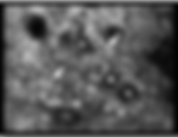 |  |
DEJ* | Basal layer | Papillary dermis |
Miguel Cordova. * Dermo-epidermal junction.
Commercial confocal microscopes for in vivo imaging:
Makes it possible to capture confocal images that depict cellular structures of living tissue (skin) in real-time. The same area of interest can be captured by the integrated dermatoscopic camera and may be imaged repeatedly over time to access both clinical and sub-clinical changes. It is being used for the bed-side evaluation of the lesion to guide patient management. The VivaScope 1500 and 3000 can be used as stand-alone units or together. Both displayed Image Resolution: 1024 x 1024 pixels However, the VivaScope 3000 offers larger field of view compared to the VivaScope 1500.
The stationary device of the VivaScope for in vivo skin imaging is designed for use on extremities such as the back of the hand or the back, chest, leg, arm, cheek or forehead.
Is a hand-held reflectance confocal microscope device for in vivo skin imaging. This imaging tool simplifies examination of difficult-to-access regions of the skin, and is particularly useful for imaging difficult locations such as lips, ears, eyes, around the nose and between fingers; all this while delivering the stable, repeatable, high quality cellular-resolution images.
Commercial confocal microscope for ex vivo imaging:
RS-G4 is a confocal microscope that is exclusively for the research purposes. But can also be used in Mohs surgery for margin assessment of the ex vivo tissue for basal cell and squamous cell carcinoma. It features a uniquely flexible scan head, innovative high-speed strip mosaic capabilities and large scale image stitching.
VivaScope 2500 is a confocal microscope specially designed for imaging fresh, needle aspirated, or fixed specimens in reflectance (phase contrast) mode or in fluorescence mode for specimens stained with fluorochromes. Specimens can be examined in near real-time without time consuming processing procedures. The VivaScope 2500 is used by physicians and other licensed healthcare professionals to view enlarged images of specimens during pathological examinations. (1)
US Centers for Medicare and Medicaid Services:
On January 1st, 2016, following more than two decades of research and development, commercialization, translational studies and clinical trials, reflectance confocal microscopy (RCM) imaging of skin was granted category I current procedural terminology (CPT) reimbursement codes (96931-96936) by the US Centers for Medicare and Medicaid Services (CMS). These codes are only applicable for in vivo reflectance confocal microscopy of skin when the examination includes mosaics at multiple levels of the epidermis and superficial dermis. Link.
Telemedicine application of RCM
Several hospitals around the world have adopted telemedicine applications for Reflectance Confocal Microscopy (RCM), also known as teleconfocal, to facilitate remote interpretation of confocal images, thereby enhancing diagnostic accuracy.
References
(3).https://jamanetwork.com/journals/jamadermatology/article-abstract/2528552
Relevant Links
Recent Developments
Handheld multiphoton and pinhole-free reflectance confocal microscopy
Real-time cross-sectional imaging of the skin, combining multiphoton microscopy (MPM) and pinhole-free reflectance confocal microscopy (RCM). The technology aims to address the limitations of traditional biopsy-based histology by providing detailed in vivo histological insights without invasive procedures.
Key features of the device include compact and lightweight design for handheld operation, making it comparable to portable ultrasound devices. It uses a Class 1M, 780 nm ultrafast pulsed NIR light source and employs advanced optical mechanisms for high-resolution imaging. The device integrates multiple optical channels to provide simultaneous imaging of various molecular and cellular features. Its cross-sectional imaging capability enables visualization of skin layers in orientations familiar to dermatologists, enhancing usability. (1)
In clinical evaluations involving 122 participants with diverse skin types (1), the system demonstrated the ability to capture histological details such as dermal elastosis, epidermal pigmentation, and pathological features of lesions like seborrheic keratosis and basal cell carcinoma.
While the system shows promise, challenges such as limited field of view, restricted imaging depth, and sensitivity to ambient light remain. Future enhancements, including integration with artificial intelligence for automated analysis and potential adaptation for endoscopic applications, could further expand its utility in dermatology and beyond (1).
Reference
Upcoming Meetings
No upcoming meetings.
Other topics

Dermoscopy
Confocal Microscopy

Diffuse Multispectral Imaging

MRI

Optical Coherence Tomography

Total Body Photography
