Total body photography (TBP) is a visual aid used by dermatologists when evaluating patients with numerous moles and who are at high risk for melanoma. Melanoma is a curable disease if diagnosed in its early stages; however, early subtle melanomas can be easy to miss. “Change” or evolution of a mole/pigmented lesion heralds a melanoma diagnosis, while the other clinical and dermoscopic features can be varied and subtle. This is the rationale behind using photography systems to map moles on the body and track them over time — it facilitates early detection of any new or changing lesions.
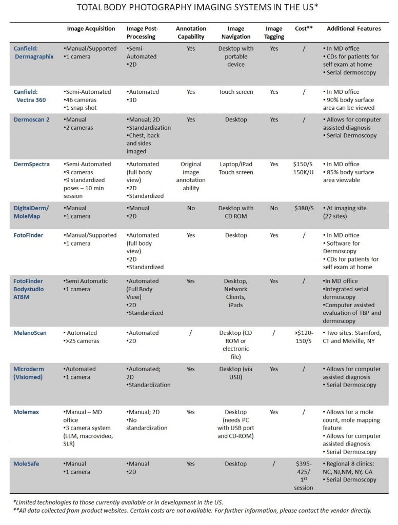
Specialized systems for image acquisition, storage, and retrieval have been developed for physicians to facilitate TBP assisted follow up and sequential digital dermoscopic monitoring. Broadly, there are imaging systems that allow for camera systems to be installed at regional sites or MD offices. These systems are typically accompanied by software that allows for total body image acquisition, storage, and retrieval with sequential tracking and advanced image standardization and comparison abilities. On the contrary, there are imaging software systems that aid dermatologists to photograph lesions with their own cameras (or a convenient handheld device from the manufacturer) to subsequently view and track standardized clinical and dermoscopic images. In addition to serving as a database for image storage and processing, total body mapping, and objective visual monitoring, most imaging software transforms a PC into a workstation for the analysis of clinical and dermoscopy pictures.. Some software also allows for computer-aided diagnosis of pigmented lesions. A comparison chart sampling a few of such systems that facilitate TBP and are available in the US market is depicted below.
The cost of TBP varies depending upon the vendor; however, it appears to be covered in case of the following parameters outlined by CMS (Medicare) under the CPT code 96904:
- A personal history of melanoma, or
- A primary blood relative (parent, sibling, or child) previously diagnosed with melanoma, or
- Multiple dysplastic nevi or atypical nevi, as determined by a clinical diagnosis performed by a pathologist or dermatologist.
In addition, certain insurances may cover this benefit if there are >50 moles or a history of immunosuppression.
To this day, there are no standards for TBP imaging that ensure the quality and interoperability of digital images. To that end, the ISIC project has been undertaken to establish a large bank of clinical and dermoscopy images, to be followed by their annotation in order to create a centralized database of images that would aid in setting up international standards for dermoscopy and total body photography.